Cellular bioelectronics
What happens to a cell culture after electrical stimulation? In E-CELL, researchers in electronics and cell biology worked together to design the chamber, deliver the stimulus and assess the response under the microscope.
Building the culture chamber
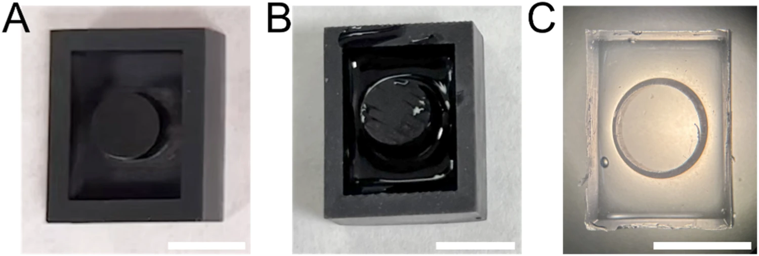
Commercial plates fix the shape and position of each well. Antonio Algarín and colleagues published a workflow for casting custom silicone wells in 3D-printed moulds and bonding them to a culture surface. They checked the assembly for leaks over 72 hours and compared short-term viability of mouse N2a cells with a standard plate in a preliminary assay.
Examining cells after stimulation
Under the direction of Alberto Yúfera and Paula Daza, E-CELL brought together all seven members of Biocircuits. One study examined mouse N2a neuroblastoma cells as monolayers and adherent neurospheres. The researchers observed more neurite-like extensions, reduced proliferation markers and less spreading of stimulated neurospheres. These are observations of early differentiation in a cell-culture model.
Can the medium carry a signal?
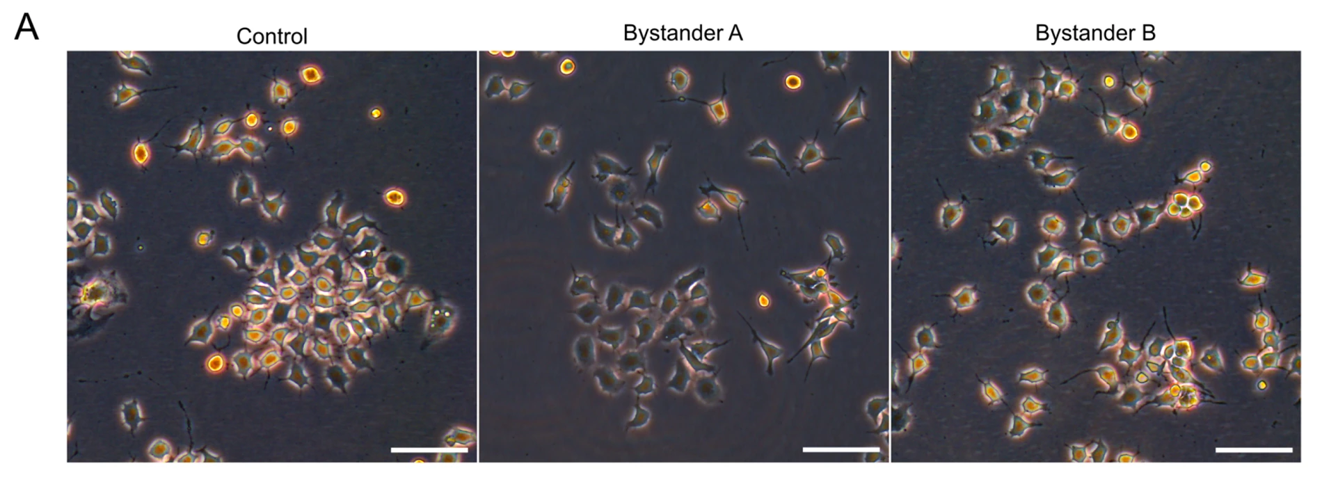
Daniel Martín and colleagues transferred medium from electrically stimulated cells to a second, unstimulated culture. The recipient cells developed longer neurites. Measurements of interleukin-6 and experiments blocking its receptor implicated this signalling pathway in the indirect response. The micrographs compare the control with cells receiving medium collected at two time points.